No content results match your keyword.
Content
You have successfully logged out.
Not registered yet?
Risk prevention
The main risks of central venous access devices misplacement include infections, bleeding, perforation of the vessel or organ, and difficulty in removing the device. Misplacement can also lead to catheter-related bloodstream infections and thrombosis, which can result in serious complications such as sepsis and pulmonary embolism. Prompt recognition and correction of misplacement are crucial to reduce these risks and improve patient outcomes.

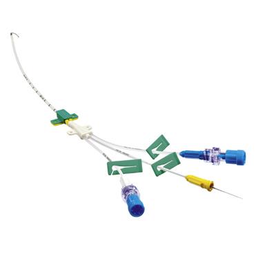
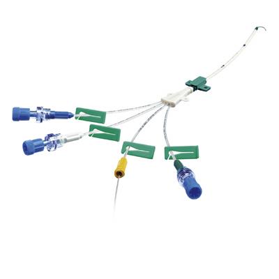
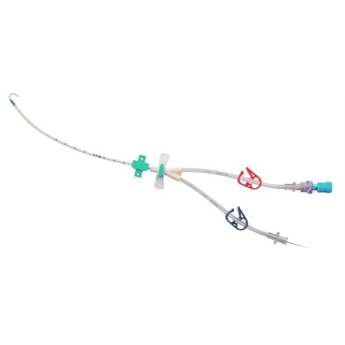
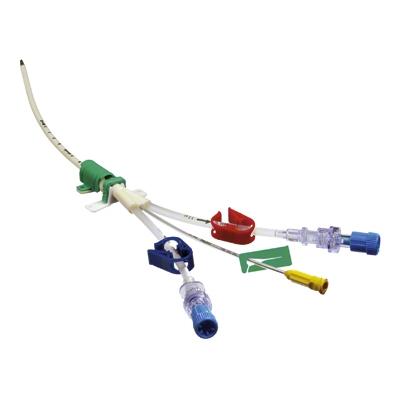
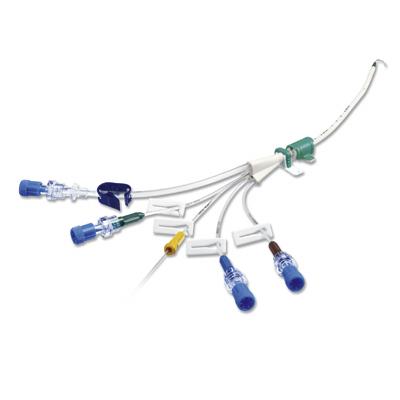
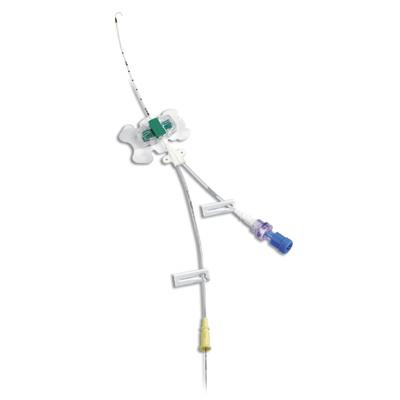
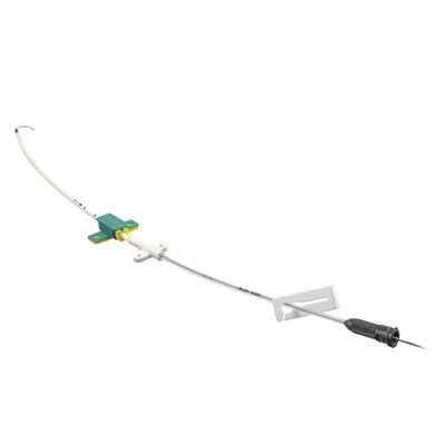
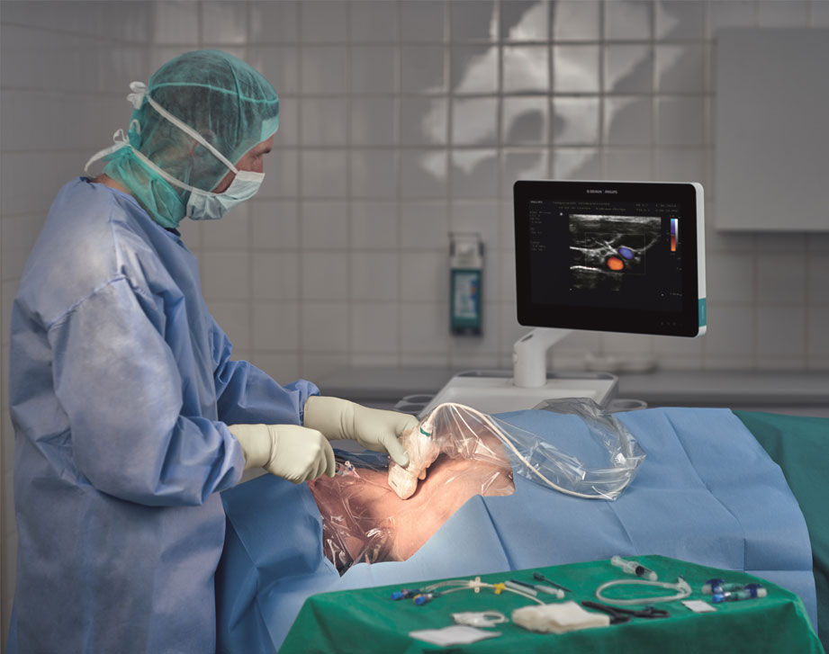
Central venous access devices (CVADs) are used for short or long-term infusion of fluids, medications and monitoring, or when establishing a peripheral venous access is not possible or difficult.CVADs can be inserted into the subclavian or jugular vein as centrally inserted central venous catheters (CICCs or conventionally called CVCs), totally implanted venous access devices (TIVAD conventionally called access ports AP), or can be inserted into one of the peripheral veins of the upper extremities, called peripherally inserted central catheters (PICCs).1
The misplacement or malposition of centrally and peripherally inserted central venous catheters describes the improper location of the catheter tip.2,3
“The tip of the catheter should be in a central vein (Superior Vena Cava, SVC, or Inferior Vena Cava, IVC), close to the cavoatrial junction.”1

„ [...] correct position of the catheter has to be ensured during placement"
 Caers J, Fontaine C,Vinh-Hung V et al (2005) Catheter tip position as a risk factor for thrombosis associated with the use of subcutaneous infusion ports. Support Care Cancer 13:325-331
Location of catheter tip position | Number of patients | Venous thrombosis | Functional problems |
| Brachocephalic vein | 32 | 45.2% | 6.5% |
| SVC cranial 1/3 | 42 | 19% | 16.7% |
| SVC mid 1/3 | 142 | 4.2% | 1.4% |
| SVC caudal 1/3 | 66 | 1.5% | 0% |
| RA or inferior Vena Cava | 18 | 5.5% | 5.6% |
Misplacement/Malposition could occur in different CVAD application

1. Right atrium
2. Caudal third of superior vena cava (SVC)
3. Cranial two-thirds of SVC or brachiocephalic veins
4. Intrathoracic part of the right subclavian vein
5. Intrathoracic part of the left subclavian vein
6. Right internal jugular vein
7. Left internal jugular vein
8. Other Graphic adapted from Pikwer A, et al. Anaesth Intensive Care. 2008; 36:30–712


Clinical needs:
*Superior vena cava thrombosis related to catheter malposition in cancer chemotherapy given through implanted ports.
.Puel V, Caudry M, Le Métayer P, Baste JC, Midy D, Marsault C, Demeaux H, Maire JP.Cancer. 1993 Oct 1;72(7):2248-52.
| Group | Thrombosis Rate |
| Catheter tip in upper SVC Left sided port | 8/28 (28.6%) |
| Catheter tip in upper SVC Right sided port | 1/33 (3%) |
| Catheter tip in lover SVC Left sided port | 0/250 |
| Catheter tip in lover SVC Right sided port | 1/68 (1.5%) |
„ […] patients with left-sided port and catheter tips lying in the upper part of the vena cava are at high risks for severe thrombotic complications.“
Support Care Cancer, 2005 May; 13(5):325-31, Epub 2004 Nov 5.
Caers J1, Fontaine C, Vinh-Hung V, De Mey J, Ponnet G, Oost C, Lamote J, De Greve J, Van Camp B, Lacor P
| Location of catheter tip | Number of patients | Venous thrombosis | Functional problems |
| Brachiocephalic vein | 31 | 45.2% | 6.5% |
| SVC cranial 1/3 | 42 | 19% | 16.7% |
| SVC mid 1/3 | 142 | 4.2% | 1.4% |
| SVC caudal 1/3 | 66 | 1.5% | 0% |
| RA or inferior Vena Cava | 18 | 5.5% | 5.6% |

Intravascular Misplacement | Extravascular Misplacement |
| Carotid artery | Extradural space |
| Azygos vein | Pericardium |
| Paersistent left sided superior vena cava | Pleural space |
| Internal thoracic (mammarx) vein | Mediastinum |
| Vertebral vein | Thoracic duct |


Clinical Consequences
| Clinical Examination and Treatment | Level of additional length of stay and cost
|
Complications related to CVAD application 1. Catheter dysfunction 2. Delay of therapy | Medica Image to detect misplaced catheter, chest X-ray, alternatively CT or MRI | + ∼ +++ |
Removal of catheter, according to the sereneness (bedside, via Interventional radiology or via surgery Insertion of new catheter. | + ∼ ++++ | |
Catheter removal via Interventional Radiology | +++ | |
Complications related to the misplaced location Carotid artery: hypotension, hemorrhagig shock Azygos vein; pleural diffusion pulmonary edema, dyspnea, chest pain, back pack pain Pericardium: fatal ventricular fibrillation | Individual surgical or non-surgicak treatment according to the impaired organ or tissue | ++ ∼ ++++ |
Drug extravasation into surrounding | Non-surgical or surgical treatment fot extravasation | ++ ∼ +++++++ |
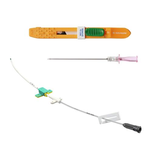
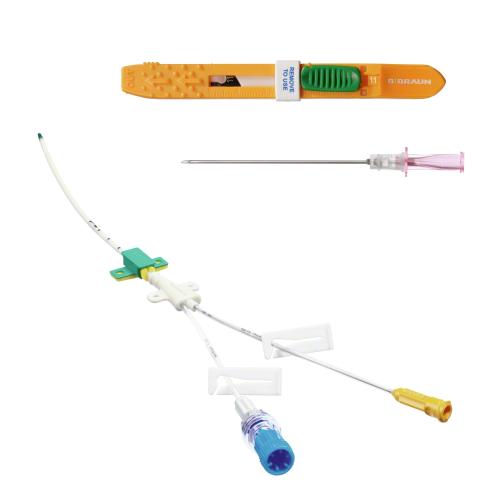
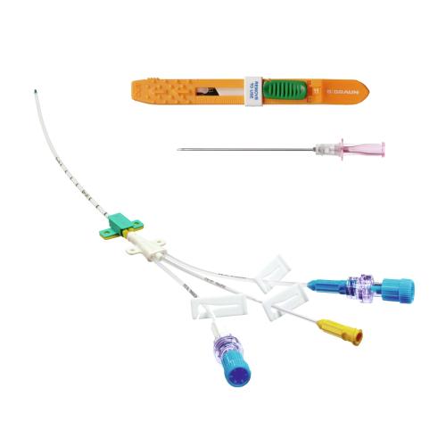
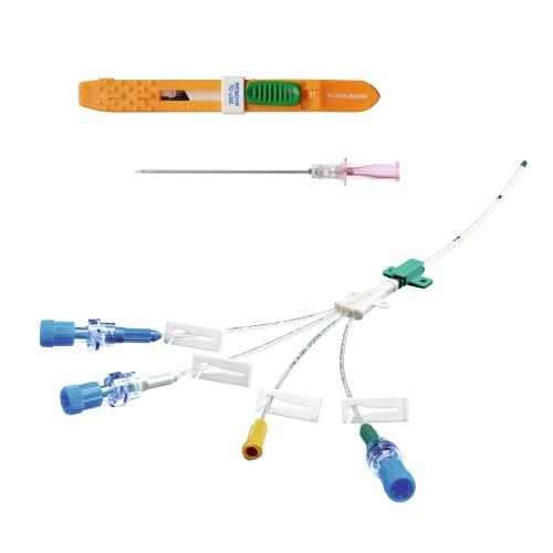
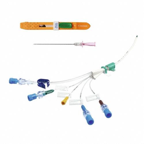
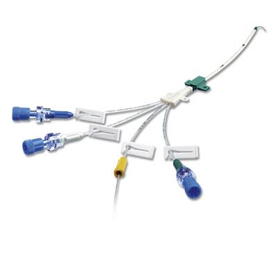
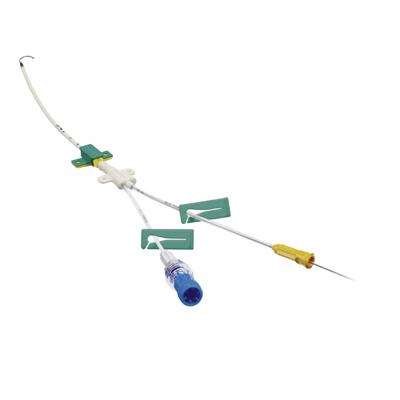
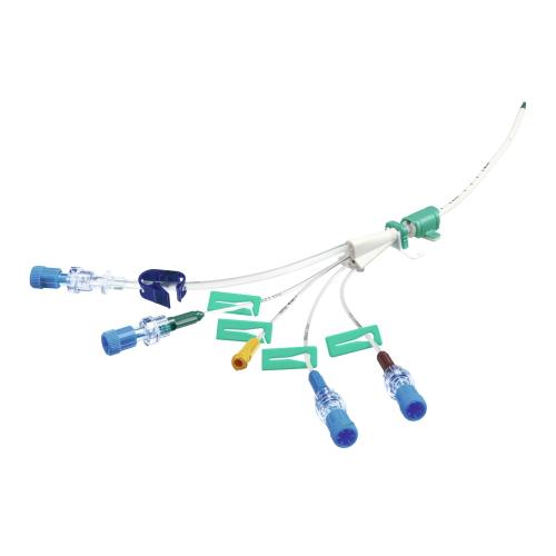
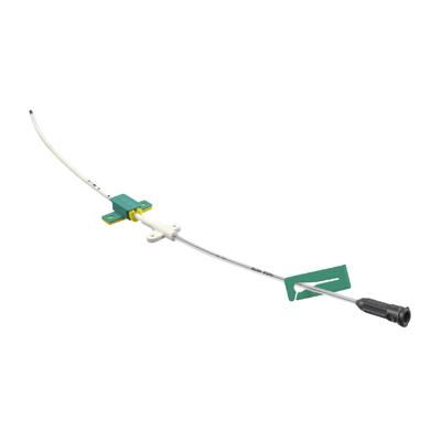
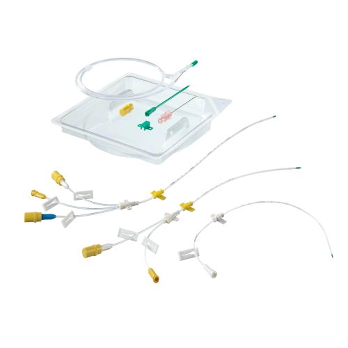
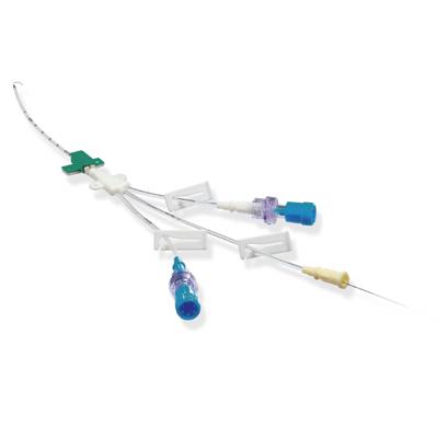
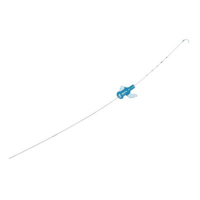
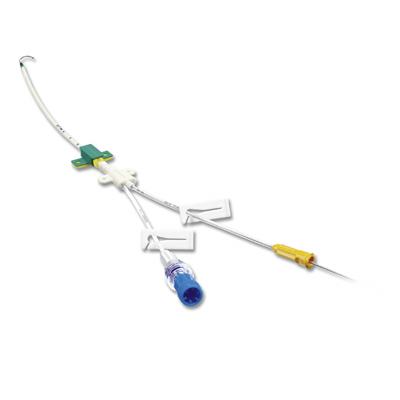
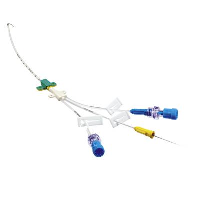
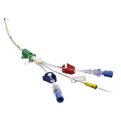
Use the different technique correctly to implant TIVADs and PICCs (Seldinger, OTW, surgical cutdown).16
Use of Valve Needles to access the vein for central venous catheters:
Save time/no disconnection needed.
Secure the CVADs sufficiently.


Adults | Children | |
| Accuracy | 94.5% | 95.8% |
| Feasibility | 98.5% | 99.3% |
Your feedback matters! Participate in our customer survey to help us enhance our website, products and services. Thank you for your support!