
Partial delivery („straddling the dura“)
Fig.2

You have successfully logged out.


Not registered yet?
Atraumatic needles for Spinal Anesthesia

Spinal Anesthesia is a trusted technique used in millions of procedures worldwide.
Yet, 5–8% of Spinal Anesthesia attempts fail1, often requiring conversion to General Anesthesia - leading to delays, discomfort, and an increased risk of complications such as post-dural puncture headache (PDPH).
Spinal Anesthesia
up to
0%
Clinical guidelines and systematic reviews consistently endorse atraumatic, pencil-point needles as the superior option for Spinal Anesthesia and lumbar puncture.3
The atraumatic, ogival tip of Pencan® separates dural fibers rather than cutting them, reducing cerebrospinal fluid (CSF) leakage and tissue damage.4
Compared to traditional cutting-tip designs, Pencan® significantly lowers the risk of post-dural puncture headache, enhances patient safety, and improves overall procedural outcomes.5
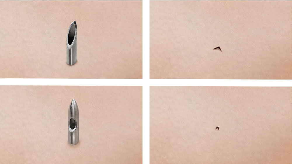 (Fig. 1) Top row pictures: Spinocan® needle with Quincke bevel and typical exertion site pattern. Bottom row pictures: Pencan® needle with pencil-point-needle and typical exertion site pattern. *Staged illustration for demonstration purposes only; not representative of actual clinical conditions.
Clinical data show that the use of Pencan® significantly reduces the incidence of post-dural puncture headache, from 7.6% with conventional Quincke needles to 2.3%. In addition, the need for an epidural blood patch is markedly reduced, decreasing from 1.8% to just 0.5% with the use of a Pencan® needle.
It is well established that atraumatic needle designs help reduce the risk of PDPH. However, atraumatic needles vary significantly between manufacturers in terms of tip geometry, orifice length, and eyelet positioning.6 Needle design should therefore be considered when selecting a needle, as differences in design features can impact performance, safety, and user experience.7
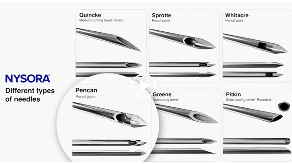
To deliver anesthetic effectively, the spinal needle must pass through several anatomical layers: skin, subcutaneous tissue, ligaments, and the dura mater. Success is confirmed by the appearance of CSF in the needle hub. However, needle design affects how reliably and safely this confirmation occurs.
Partial placement can occur, where the orifice lies between the subarachnoid and epidural spaces. This can lead to incomplete drug delivery and therefore might increase the likelihood of failed blocks (Fig. 2). Small orifices geometrically are less likely to open in both spaces.8
Placing a needle too deep into the spinal space might increase the risk of nerve contact and the chance of paresthesia9 (Fig. 3). A shorter tip-to-back-of-eyelet distance could help minimize the insertion depth of the needle tip into the subarachnoid space. It can also enable faster CSF return for better control during placement.
Fig.2

Fig. 3
Designed to support full intrathecal delivery to reduce the likelihood of partial or incomplete anesthesia.
Eyelet positioning is designed for faster CSF flashback, which could help with correct and quicker placement of the needle.
Provides a clear “dural pop” for tactile feedback, helps minimize nerve contact, and promotes a clean dural tear—associated with lower PDPH rates.
In the tight spinal space the risk of cauda equina contact should be avoided, especially in pediatric patients. A short orifice to tip distance needs less penetration depth in the spinal space.
WHO recommendations
Intrapartum care for a positive childbirth experience - WHO recommendations: intrapartum care for a positive childbirth experience
link
WFSA Labour Analgesia Declaration
Labour Analgesia Declaration – WFSA
link
ASA Practice Guidelines for Obstetric Anesthesia
An Updated Report by the American Society of Anesthesiologists Task Force on Obstetric Anesthesia and the Society for Obstetric Anesthesia and Perinatology Anesthesiology
link
ASA Statement on Neuraxial Analgesia or Anesthesia in Obstetrics
Standards and Practice Parameters Statement on Neuraxial Anesthesia in Obstetrics
link
A responsible contact person will get in touch with you as soon as possible.
1. Kinsella, S.M. (2008). A prospective audit of regional anaesthesia failure in 5080 Caesarean sections. Anaesthesia, 63(8), 822–832. doi: 10.1111/j.1365-2044.2008.05499.x.
2. Batova, R., & Georgiev, S. (2019). Impact of spinal needle design and approach on postdural puncture headache and spinal anesthesia failure in obstetrics. Anaesthesiology Intensive Therapy, 51(2), 77–82. DOI: 10.5114/ait.2019.86166
3. Nath, S., Koziarz, A., Badhiwala, J. H., et al. (2018). Atraumatic versus conventional lumbar puncture needles: A systematic review and meta-analysis. The Lancet, 391(10126), 1197–1204. https://doi.org/10.1016/S0140-6736(17)32451-0
4. Reina, M. A., Puigdellívol-Sánchez, A., Gatt, S. P., De Andrés, J. A., Prats-Galino, A., & van Zundert, A. (2017). Electron microscopy of dural and arachnoid disruptions after subarachnoid block. Regional Anesthesia and Pain Medicine, 42(6), 709–718. https://doi.org/10.1097/AAP.0000000000000667
5. Xu H, Liu Y, Song W, Kan S, Liu F, Zhang D, Ning G, Feng S. Comparison of cutting and pencil-point spinal needle in spinal anesthesia regarding postdural puncture headache: A meta-analysis. Medicine (Baltimore). 2017 Apr;96(14):e6527. doi: 10.1097/MD.0000000000006527. PMID: 28383416; PMCID: PMC5411200.
6. B. Braun Melsungen AG internal technical data report for Spinal needles (2025); as well as manufacturer’s information provided by Pajunk GmbH (2026).
7. Reina, M. A., López, A., De Andrés, J. A., Prats-Galino, A., & van Zundert, A. (2007). Variability in the design of spinal needles: Implications for clinical practice. Regional Anesthesia and Pain Medicine, 32(4), 346–353. https://doi.org/10.1016/j.rapm.2007.02.006
8. Rae, J. D., & Fettes, P. D. W. (2023). Mechanisms and management of failed spinal anesthesia. NYSORA – The New York School of Regional Anesthesia. Retrieved from https://www.nysora.com/topics/complications/mechanisms-management-failed-spinal-anesthesia/
9. Reina MA, De Andres JA, Hernández JM, Arriazu Navarro R, Pastor J, Prats-Galino A. Looking for the development of paresthesias in the subarachnoid and epidural anesthesia: A clinical and anatomical analysis. Reg Anesth Pain Med. 2011;36(Suppl):E17–E22. doi:10.1097/AAP.0b013e3182030648. https://www.researchgate.net/publication/265377231_LOOKING_FOR_THE_DEVELOPMENT_OF_PARESTHESIAS_IN_THE_SUBARACHNOID_AND_EPIDURAL_ANAESTHESIA_A_CLINICAL_AND_ANATOMICAL_ANALYSIS_MA_Reina
Your feedback matters! Participate in our customer survey to help us enhance our website, products and services. Thank you for your support!


