No content results match your keyword.
Content
You have successfully logged out.
Not registered yet?
Help prevent and treat diabetic foot ulcers
Diabetic foot syndrome (DFS) is a common complication of diabetes mellitus that affects every fourth patient with diabetes at some point in their lifes. The risk of developing DFS increases with age.
One of the consequences is skin breakdown or foot ulceration, known as a Diabetic Foot Ulcer (DFU). This condition includes all changes in the foot resulting from diabetic polyneuropathy as well as diabetic micro and macroangiopathic changes.

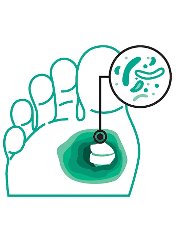
Infection in Diabetic Foot Ulcers (DFU)
19- 0%
of people with diabetes will develop a DFU during their lifetime.[2]
More than
0%
of such diabetic foot ulcers become infected.[3]
Up to
0%
of severely infected DFUs lead to osteomyelitis.[4]
Diabetic Foot Ulcers Infection: Prevention & Treatment
Infection in DFU is associated not only with poor or delayed healing but also poses a risk factor for hospitalization. Furthermore, 10% to 45% of patients hospitalized for foot infection require readmission within one year, leading to increased healthcare costs. In individuals with severe infection or osteomyelitis, the probability of amputation rises significantly.[5]
Diagnosis of DFU is mainly based on clinical signs:[5]
And clinical examination techniques:[6]
The severity of diabetic foot infections is classified according to the IWGDF/IDSA classification:[6]
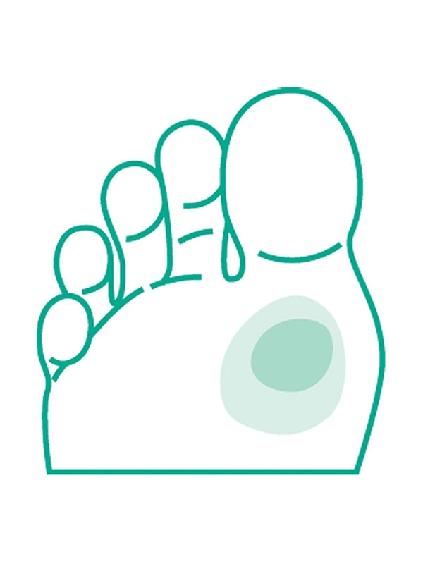
No systemic or local symptoms or signs of infection.
Cleanse with wound cleansing solutions and debride.
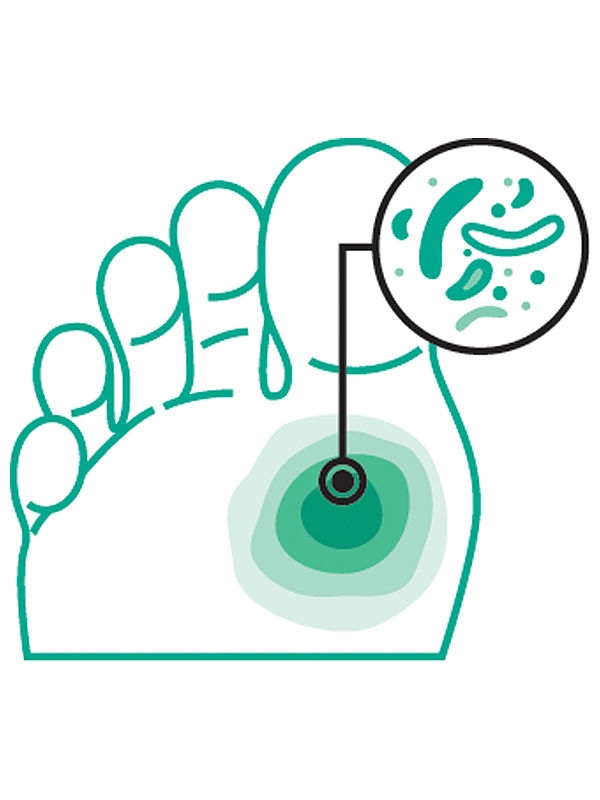
Infected: At least two of these conditions are present: local swelling or induration; erythema >0.5 but <2 cm around the wound; local tenderness or pain; local increased warmth; purulent discharge.
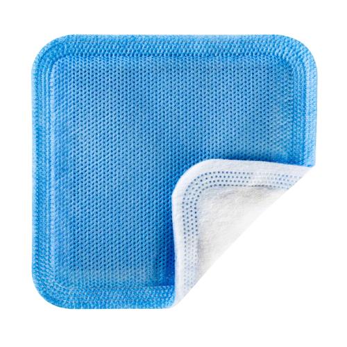
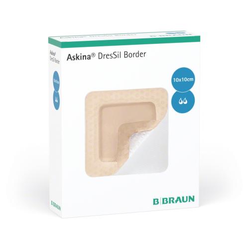
Cleanse with wound cleansing solutions, debride and apply silver dressings to control local infection. Treat with systemic antibiotics (oral agents).
add “O” if osteomyelitis is present
Infection with no systemic manifestations. Involving: erythema extending ≥2 cm from the wound margin, and/or affected tissue deeper than skin and subcutaneous tissues (e.g., tendon, muscle, joint, and bone).
Refer patient to multidisciplinary diabetic foot care teams. Treat with systemic antibiotics (oral/parenteral). Surgical debridement if it is needed.
add “O” if osteomyelitis is present
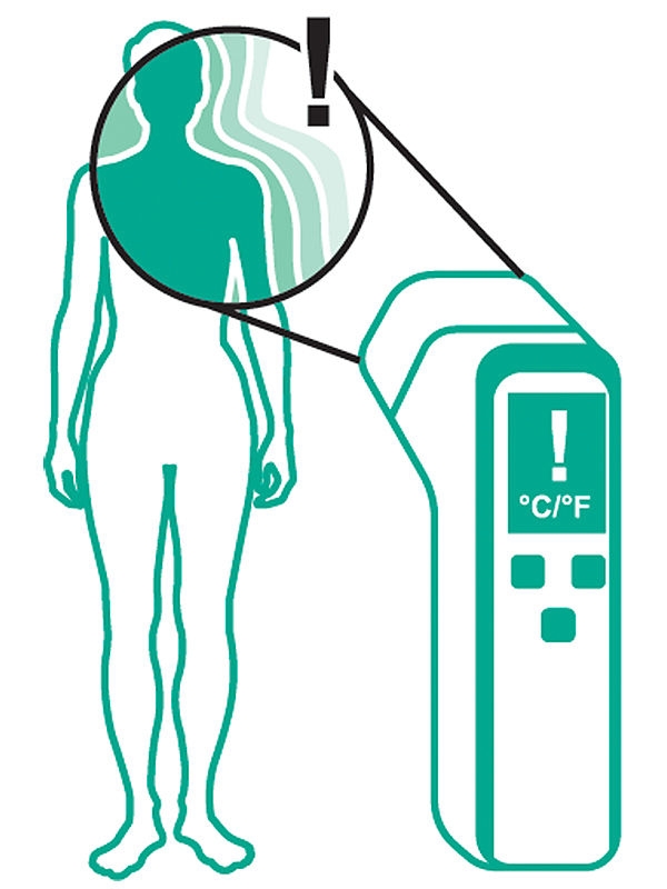
Any foot infection ≥2 cm with associated systemic manifestations, such as: temperature >38°C or <36°C; heart rate >90 beats/min; respiratory rate >20 breaths/min or PaCO2 < 4.3 kPa (32 mmHg); white blood cell count >12,000/mm3 or < 4 G/L, or >10% immature (band) forms.
Refer to multidisciplinary diabetic foot care teams. Treat with systemic antibiotics (parenteral). Surgical debridement if it is needed.
Biofilm is present in more than 78% of diabetic foot ulcers.[8]
If biofilm is suspected in a diabetic foot ulcer, antibiofilm strategies should be established with the dual objective of helping to prevent the development of an infection and helping to promote ulcer healing.[7]
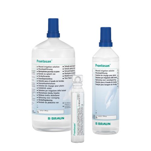
Clean the wound using cleansing solutions that contain surfactant agents to help break down and remove biofilm. Solutions containing polyhexanide and betaine have shown beneficial effects in disrupting biofilm and improving wound pH. It is recommended to apply the cleansing solution using soaked compresses for approximately 5 minutes. Follow this with gentle brushing of the wound bed to rinse the area thoroughly and prepare it for debridement of non-viable tissue.
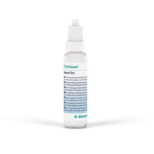
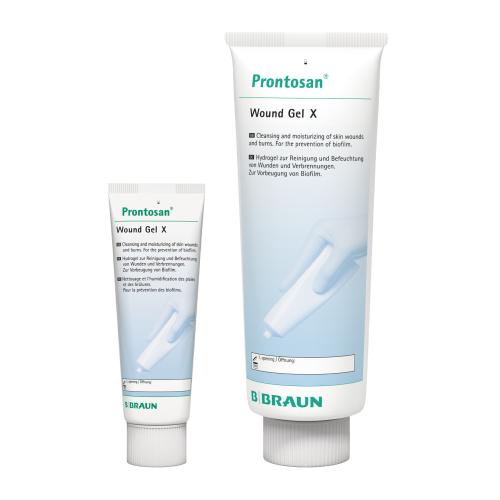
Sharp debridement is the preferred method for treating diabetic foot ulcers. This involves the use of a scalpel, scissors, or curette to remove necrotic tissue until slight bleeding is observed, indicating viable tissue. The effectiveness of this procedure is enhanced by prior application of cleansing agents such as Prontosan®.
The use of silver dressings prevents biofilm reformation and reduces bacterial load.[7]
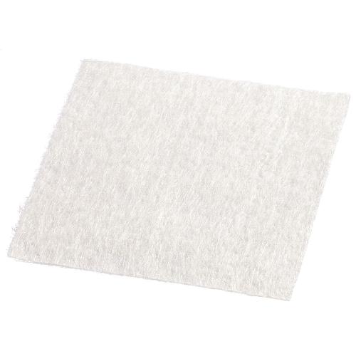
Use suitable dressings such as alginate or superabsorbent wound dressings for effective control of wound exudate. Excessive moisture can lead to maceration of surrounding skin and delay healing.[7]
Your feedback matters! Participate in our customer survey to help us enhance our website, products and services. Thank you for your support!