No content results match your keyword.
Content
You have successfully logged out.
Not registered yet?
AESCULAP® EinsteinVision® 3.0 FI – SEE BETTER. SEE BEYOND.
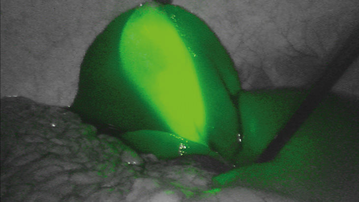
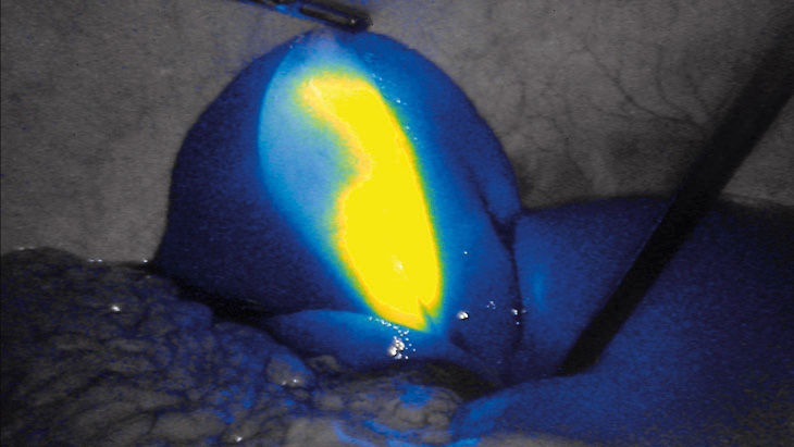
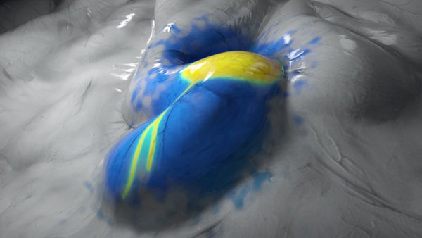
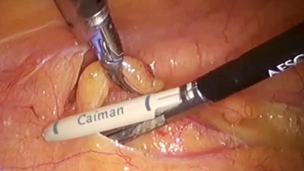
Fluorescence Imaging (FI) aims for the best patient outcome during diagnostics or surgery. [1] In minimally invasive surgery it is used in numerous applications such as vessel or visceral perfusion assessment, visualization of bile duct anatomy or (sentinel) lymph node mapping.

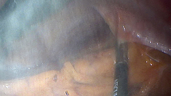
Integrated heating elements in the endoscope tip of the camera effectively and permanently prevent fogging of the optics within seconds.


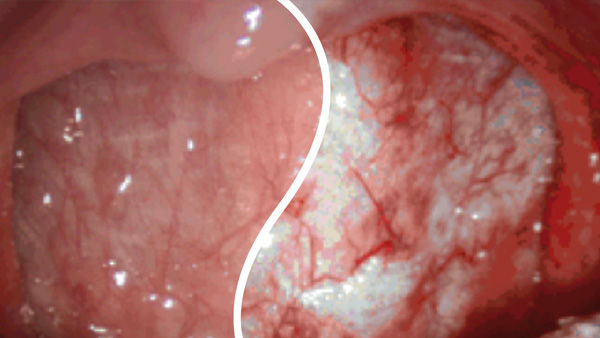
With the push of a button, a special algorithm reduces the impairment visibly.
The integrated algorithm provides precisely that at the push of a button.
Dr. med. Mirko Otto, Acting Senior Physician, Surgical Clinic, Mannheim University Medical Center
Read moreDr. med. Bernd Heinzmann, Senior Physician, Surgery/Pediatric Surgery, St. Marienstift Hospital Magdeburg GmbH
Read morePriv.-Doz. Dr. med. Peter Grimminger, Senior Consultant Head Upper GI Surgery, Minimally Invasive Surgery and Robotics, University Medical Center of the Johannes Gutenberg University Mainz
Read moreDr. med. Max Mayr, Senior Physician, Clinic for General and Visceral Surgery Barmherzigen Brüder Regensburg Hospital
Prof. Dr. Jörg Glatzle, Senior Consultant of the Clinic for General and Visceral Surgery Deputy Medical Director, Konstanz Hospital
Could fluorescence-guided surgery be an efficient and sustainable option?
link
Compendium on AESCULAP® EinsteinVision® 3.0 FI
link
Brochure EinsteinVision® 3.0 FI
link
Brochure EinsteinVision® 3.0
link
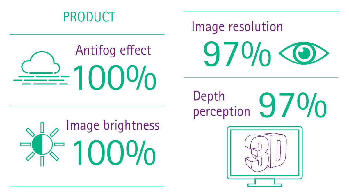
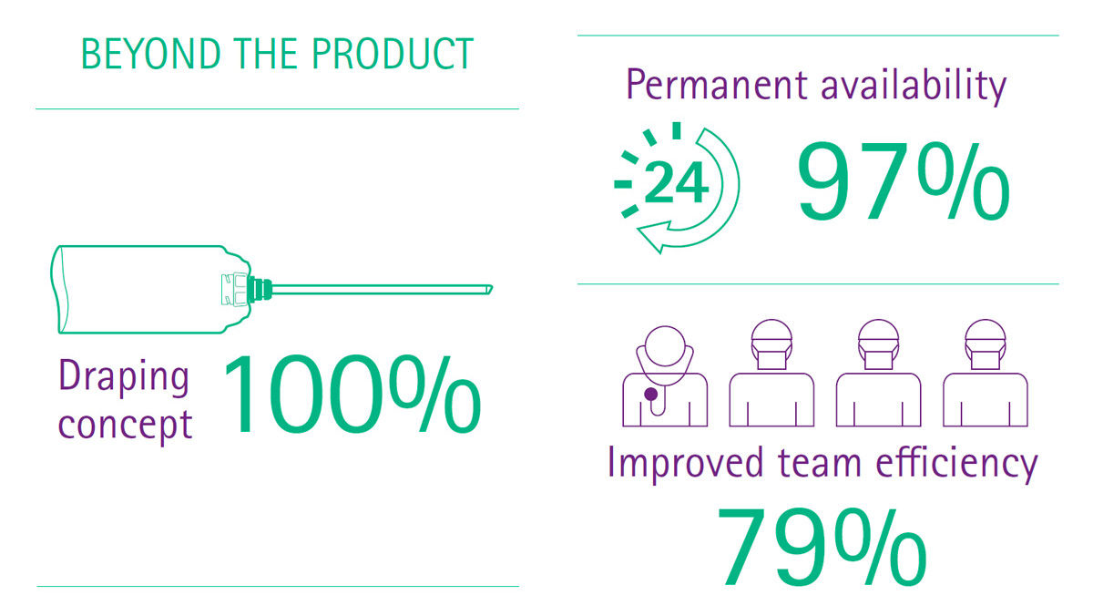
Info graph proven value of 3D EinsteinVision® 3.0 FI
link
Info graph proven value of 3D EinsteinVision® 3.0
link
Medical Technologies from B. Braun, such as EinsteinVision®, assist your surgical team in its daily practice. So that you can focus on providing medical care and not on cost management, we offer customized financing solutions that adapt flexibly to your institution’s needs. Download for more information.
Download for more informationReferences
Your feedback matters! Participate in our customer survey to help us enhance our website, products and services. Thank you for your support!
